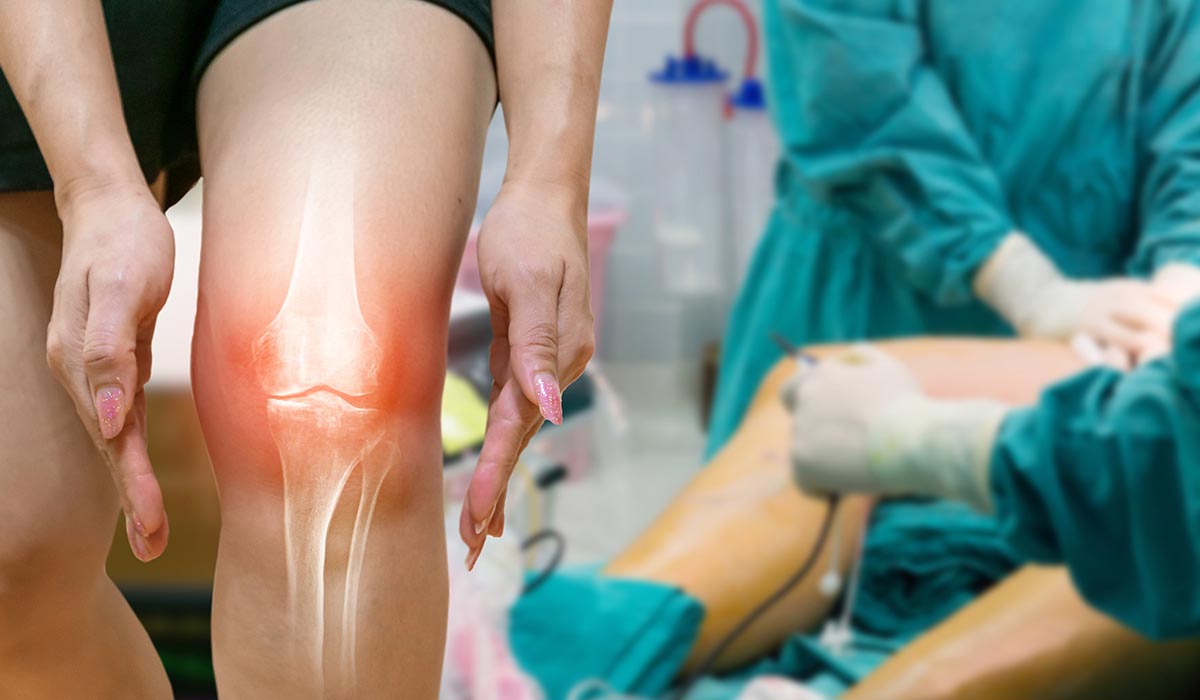
Joint replacement surgery, such as Total Hip Replacement (THR) and Total Knee Replacement (TKR), has become a routine procedure worldwide, offering relief to patients suffering from severe arthritis, joint trauma, or degenerative joint diseases. These surgeries significantly improve a patient’s quality of life by reducing pain, improving mobility, and restoring joint function. However, in some cases, a previously successful joint replacement may fail over time, necessitating a Revision Joint Replacement Surgery.
Revision joint replacement is a complex surgical procedure where an existing prosthesis (artificial joint implant) is replaced due to failure, complications, or other medical reasons. Unlike primary joint replacement surgery, which focuses on replacing a damaged joint, revision surgery addresses problems related to the previous implant, bone, and surrounding tissues.
Understanding the indications for revision joint replacement surgery is essential for patients, caregivers, and healthcare professionals to ensure timely and appropriate intervention. Below, we explore in detail the various medical reasons that necessitate revision joint replacement surgery.
1. Periprosthetic Joint Infection (PJI) – The Most Common Indication
What is Periprosthetic Joint Infection?
A Periprosthetic Joint Infection (PJI) occurs when bacteria infect the area around the artificial joint. It is regarded as the most common and serious reason for revision joint replacement surgery. Infections can be classified based on their time of onset:
- Early infections – Occur within weeks after the primary surgery.
- Delayed infections – Occur months or years after the initial procedure.
- Late infections – Result from hematogenous spread, where bacteria from another infection site (e.g., urinary tract infection) reach the implant.
How Does Infection Cause Implant Failure?
Bacteria form biofilms around the prosthetic surface, making them resistant to antibiotics and the body’s immune defenses. This leads to:
- Persistent inflammation
- Severe pain
- Implant loosening
- Loss of joint function
If untreated, the infection can spread to surrounding bones and tissues, significantly worsening the patient’s condition.
Clinical Symptoms
- Swelling and redness around the joint
- Increased warmth
- Persistent joint pain
- Fever
- Drainage of pus from the surgical site in some cases
Diagnosis of Infection
- Blood tests: Elevated Erythrocyte Sedimentation Rate (ESR) and C-Reactive Protein (CRP) levels indicate inflammation.
- Joint aspiration and culture: Helps detect and identify bacteria responsible for infection.
- Imaging studies: X-rays, MRI, or nuclear scans help visualize implant loosening or bone destruction.
Treatment Protocol
Most cases of PJI require a two-stage revision approach:
- Stage 1:
– Removal of the infected prosthesis
– Thorough debridement (removal of infected tissue)
– Placement of an antibiotic-impregnated spacer to maintain joint space and deliver high local antibiotic concentration - Stage 2:
– After 6–12 weeks and confirmation of infection eradication, a new prosthesis is implanted.
In selected cases, a single-stage revision surgery is possible when the infection is detected early and the patient is fit, which reduces hospital stay and overall cost.
2. Aseptic Loosening of Implants
What is Aseptic Loosening?
Aseptic loosening is defined as the gradual failure of the bond between the implant and the surrounding bone without the presence of infection. It is the second most common cause of revision joint replacement surgeries.
Why Does Aseptic Loosening Occur?
Several factors contribute to aseptic loosening:
- Osteoporosis: Reduced bone density weakens the bone-implant interface, leading to gradual loosening.
- Insufficient Cement Mantle: In cases where the prosthesis is fixed with bone cement, poor cement technique or insufficient coverage can result in poor fixation.
- Inappropriate Implant Size or Design: A prosthesis that is too small, too large, or improperly designed can cause abnormal joint biomechanics, accelerating loosening.
- Wear Particle Induced Osteolysis: Microscopic particles shed from polyethylene or metal components stimulate an inflammatory response, which activates osteoclasts (bone-resorbing cells), leading to bone loss around the implant.
Symptoms
- Chronic joint pain
- Joint instability
- Decreased range of motion
- Difficulty bearing weight on the affected limb
Diagnosis
- X-rays: Show radiolucent lines between the implant and bone, implant migration, or bone loss.
- Bone Scans: Detect areas of increased metabolic activity, indicating loosening or bone remodeling.
Treatment Approach
- Removal of the loose prosthesis
- Bone grafting (if necessary) to restore bone stock
- Insertion of a new implant, often using a long-stemmed design or specialized modular implants that offer better fixation and load distribution.
3. Periprosthetic Fractures
What Are Periprosthetic Fractures?
A periprosthetic fracture is a break in the bone surrounding an existing prosthesis. These fractures occur either due to trauma (e.g., a fall) or weakening of bone over time, especially in osteoporotic patients.
Why Is Revision Necessary?
- A periprosthetic fracture can destabilize the implant, resulting in pain, deformity, and loss of function.
- In some cases, simple fracture fixation is insufficient due to poor bone quality or implant instability, necessitating a full revision surgery.
Common Locations
- Femur (around hip prosthesis)
- Tibia or femur (around knee prosthesis)
Diagnosis
- X-rays showing fracture lines around the implant
- CT scans for detailed assessment of bone defects and fracture pattern
Treatment Approach
- Stable Implant + Minor Fracture → Fracture fixation using plates and screws.
- Unstable Implant or Severe Fracture → Removal of the old prosthesis and replacement with a longer-stemmed revision implant that bridges the fracture.
- In cases of massive bone loss, structural allografts (bone from a donor) or specialized implant designs are used.
4. Implant Malposition and Joint Instability
Causes of Malposition
- Technical errors during the initial surgery
- Postoperative implant migration over time
- Inadequate soft tissue balancing
Symptoms
- Joint instability (clicking or sensation of the joint giving way)
- Limb length discrepancy
- Abnormal gait pattern
- Persistent pain
Treatment Approach
- Removal of malpositioned implant
- Accurate placement of a new prosthesis with correct alignment
- Soft tissue reconstruction when necessary (e.g., ligament repair).
5. Mechanical Wear and Implant Failure
Why Does Mechanical Wear Occur?
Artificial joint components are subject to mechanical wear over time due to repeated friction during movement. This leads to:
- Wear of polyethylene liners
- Metal corrosion
- Microparticle shedding
These wear particles cause chronic inflammation, osteolysis, and loosening of the implant.
Symptoms
- Progressive joint pain
- Decreased function
- Swelling or warmth in the joint
Treatment Approach
- Removal of worn components
- Replacement with advanced implants made of highly cross-linked polyethylene, ceramic materials, or cobalt-chromium alloys designed to minimize wear.
6. Severe Bone Loss (Bone Deficiency)
What Causes Bone Loss?
- Long-standing implant failure
- Osteolysis from wear particles
- Infection
- Trauma
Why Is Bone Loss a Concern?
Significant bone loss compromises implant stability and may prevent adequate fixation of a new prosthesis.
Treatment Approach
- Bone grafting (autograft or allograft) to restore bone stock.
- Use of modular or custom implants with longer stems to provide enhanced fixation.
- Specialized implants with porous surfaces to encourage bone ingrowth.
7. Allergic Reactions to Implant Materials
Although rare, some patients develop allergic reactions to the metals used in implants (e.g., nickel, cobalt, chromium). This causes:
- Chronic joint pain
- Skin rashes
- Localized swelling
- Persistent inflammation
Treatment Approach
- Removal of the allergenic implant
- Replacement with hypoallergenic implants made of titanium or ceramic materials, which are biocompatible and less likely to cause allergic reactions.
Challenges of Revision Joint Replacement Surgery
Revision joint replacement is inherently more complicated than primary replacement due to:
- Extensive scar tissue and poor bone stock
- Higher risk of infection
- Increased operative time and blood loss
- Need for specialized implants and surgical techniques
However, modern advancements such as robotic-assisted surgery, 3D preoperative planning, and patient-specific implants have significantly improved outcomes and reduced complications.
Postoperative Care and Recovery
After revision surgery, patients require:
- Close monitoring for early signs of infection or implant failure.
- Pain management protocols tailored to individual needs.
- Physiotherapy to help restore joint function gradually.
- Nutritional support, particularly calcium and vitamin D, to promote bone healing.
- Regular follow-up visits with imaging studies to ensure proper implant positioning and stability.
Our experienced team at ORTHO D HOSPITAL offers a customized postoperative rehabilitation plan to help patients recover as quickly and safely as possible, returning them to a functional and pain-free life.
Conclusion
Revision joint replacement surgeries play a critical role in restoring joint function and improving quality of life when a previous joint replacement fails due to:
- Periprosthetic joint infection
- Aseptic loosening
- Periprosthetic fractures
- Implant malposition or instability
- Mechanical wear and implant failure
- Bone loss
- Allergic reactions to implant materials
These surgeries require advanced planning, specialized implants, and an experienced surgical team. At ORTHO D HOSPITAL, we provide comprehensive care for NRI and international patients seeking high-quality revision joint replacement solutions in India.
Our mission is to offer advanced surgical solutions, compassionate care, and long-term functional restoration for patients facing joint replacement failure.