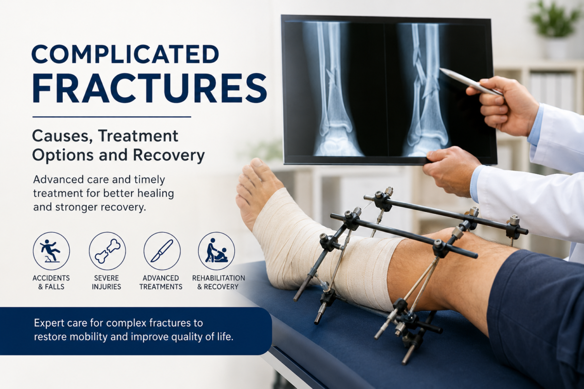
What Are the Indications for Revision Joint Replacement Surgeries?

Joint replacement surgery, such as Total Hip Replacement (THR) and Total Knee Replacement (TKR), has become a routine procedure worldwide, offering relief to patients suffering from severe arthritis, joint trauma, or degenerative joint diseases. These surgeries significantly improve a patient’s quality of life by reducing pain, improving mobility, and restoring joint function. However, in some cases, a previously successful joint replacement may fail over time, necessitating a Revision Joint Replacement Surgery.

Revision joint replacement is a complex surgical procedure where an existing prosthesis (artificial joint implant) is replaced due to failure, complications, or other medical reasons. Unlike primary joint replacement surgery, which focuses on replacing a damaged joint, revision surgery addresses problems related to the previous implant, bone, and surrounding tissues.

Understanding the indications for revision joint replacement surgery is essential for patients, caregivers, and healthcare professionals to ensure timely and appropriate intervention. Below, we explore in detail the various medical reasons that necessitate revision joint replacement surgery.

1. Periprosthetic Joint Infection (PJI) – The Most Common Indication

What is Periprosthetic Joint Infection?

A Periprosthetic Joint Infection (PJI) occurs when bacteria infect the area around the artificial joint. It is regarded as the most common and serious reason for revision joint replacement surgery. Infections can be classified based on their time of onset:

  • Early infections – Occur within weeks after the primary surgery.
  • Delayed infections – Occur months or years after the initial procedure.
  • Late infections – Result from hematogenous spread, where bacteria from another infection site (e.g., urinary tract infection) reach the implant.

How Does Infection Cause Implant Failure?

Bacteria form biofilms around the prosthetic surface, making them resistant to antibiotics and the body’s immune defenses. This leads to:

  • Persistent inflammation
  • Severe pain
  • Implant loosening
  • Loss of joint function

If untreated, the infection can spread to surrounding bones and tissues, significantly worsening the patient’s condition.

Clinical Symptoms

  • Swelling and redness around the joint
  • Increased warmth
  • Persistent joint pain
  • Fever
  • Drainage of pus from the surgical site in some cases

Diagnosis of Infection

  • Blood tests: Elevated Erythrocyte Sedimentation Rate (ESR) and C-Reactive Protein (CRP) levels indicate inflammation.
  • Joint aspiration and culture: Helps detect and identify bacteria responsible for infection.
  • Imaging studies: X-rays, MRI, or nuclear scans help visualize implant loosening or bone destruction.

Treatment Protocol

Most cases of PJI require a two-stage revision approach:

  • Stage 1:
    – Removal of the infected prosthesis
    – Thorough debridement (removal of infected tissue)
    – Placement of an antibiotic-impregnated spacer to maintain joint space and deliver high local antibiotic concentration
  • Stage 2:
    – After 6–12 weeks and confirmation of infection eradication, a new prosthesis is implanted.

In selected cases, a single-stage revision surgery is possible when the infection is detected early and the patient is fit, which reduces hospital stay and overall cost.

2. Aseptic Loosening of Implants

What is Aseptic Loosening?

Aseptic loosening is defined as the gradual failure of the bond between the implant and the surrounding bone without the presence of infection. It is the second most common cause of revision joint replacement surgeries.

Why Does Aseptic Loosening Occur?

Several factors contribute to aseptic loosening:

  • Osteoporosis: Reduced bone density weakens the bone-implant interface, leading to gradual loosening.
  • Insufficient Cement Mantle: In cases where the prosthesis is fixed with bone cement, poor cement technique or insufficient coverage can result in poor fixation.
  • Inappropriate Implant Size or Design: A prosthesis that is too small, too large, or improperly designed can cause abnormal joint biomechanics, accelerating loosening.
  • Wear Particle Induced Osteolysis: Microscopic particles shed from polyethylene or metal components stimulate an inflammatory response, which activates osteoclasts (bone-resorbing cells), leading to bone loss around the implant.

Symptoms

  • Chronic joint pain
  • Joint instability
  • Decreased range of motion
  • Difficulty bearing weight on the affected limb

Diagnosis

  • X-rays: Show radiolucent lines between the implant and bone, implant migration, or bone loss.
  • Bone Scans: Detect areas of increased metabolic activity, indicating loosening or bone remodeling.

Treatment Approach

  • Removal of the loose prosthesis
  • Bone grafting (if necessary) to restore bone stock
  • Insertion of a new implant, often using a long-stemmed design or specialized modular implants that offer better fixation and load distribution.

3. Periprosthetic Fractures

What Are Periprosthetic Fractures?

A periprosthetic fracture is a break in the bone surrounding an existing prosthesis. These fractures occur either due to trauma (e.g., a fall) or weakening of bone over time, especially in osteoporotic patients.

Why Is Revision Necessary?

  • A periprosthetic fracture can destabilize the implant, resulting in pain, deformity, and loss of function.
  • In some cases, simple fracture fixation is insufficient due to poor bone quality or implant instability, necessitating a full revision surgery.

Common Locations

  • Femur (around hip prosthesis)
  • Tibia or femur (around knee prosthesis)

Diagnosis

  • X-rays showing fracture lines around the implant
  • CT scans for detailed assessment of bone defects and fracture pattern

Treatment Approach

  • Stable Implant + Minor Fracture → Fracture fixation using plates and screws.
  • Unstable Implant or Severe Fracture → Removal of the old prosthesis and replacement with a longer-stemmed revision implant that bridges the fracture.
  • In cases of massive bone loss, structural allografts (bone from a donor) or specialized implant designs are used.

4. Implant Malposition and Joint Instability

Causes of Malposition

  • Technical errors during the initial surgery
  • Postoperative implant migration over time
  • Inadequate soft tissue balancing

Symptoms

  • Joint instability (clicking or sensation of the joint giving way)
  • Limb length discrepancy
  • Abnormal gait pattern
  • Persistent pain

Treatment Approach

  • Removal of malpositioned implant
  • Accurate placement of a new prosthesis with correct alignment
  • Soft tissue reconstruction when necessary (e.g., ligament repair).

5. Mechanical Wear and Implant Failure

Why Does Mechanical Wear Occur?

Artificial joint components are subject to mechanical wear over time due to repeated friction during movement. This leads to:

  • Wear of polyethylene liners
  • Metal corrosion
  • Microparticle shedding

These wear particles cause chronic inflammation, osteolysis, and loosening of the implant.

Symptoms

  • Progressive joint pain
  • Decreased function
  • Swelling or warmth in the joint

Treatment Approach

  • Removal of worn components
  • Replacement with advanced implants made of highly cross-linked polyethylene, ceramic materials, or cobalt-chromium alloys designed to minimize wear.

6. Severe Bone Loss (Bone Deficiency)

What Causes Bone Loss?

  • Long-standing implant failure
  • Osteolysis from wear particles
  • Infection
  • Trauma

Why Is Bone Loss a Concern?

Significant bone loss compromises implant stability and may prevent adequate fixation of a new prosthesis.

Treatment Approach

  • Bone grafting (autograft or allograft) to restore bone stock.
  • Use of modular or custom implants with longer stems to provide enhanced fixation.
  • Specialized implants with porous surfaces to encourage bone ingrowth.

7. Allergic Reactions to Implant Materials

Although rare, some patients develop allergic reactions to the metals used in implants (e.g., nickel, cobalt, chromium). This causes:

  • Chronic joint pain
  • Skin rashes
  • Localized swelling
  • Persistent inflammation

Treatment Approach

  • Removal of the allergenic implant
  • Replacement with hypoallergenic implants made of titanium or ceramic materials, which are biocompatible and less likely to cause allergic reactions.

Challenges of Revision Joint Replacement Surgery

Revision joint replacement is inherently more complicated than primary replacement due to:

  • Extensive scar tissue and poor bone stock
  • Higher risk of infection
  • Increased operative time and blood loss
  • Need for specialized implants and surgical techniques

However, modern advancements such as robotic-assisted surgery, 3D preoperative planning, and patient-specific implants have significantly improved outcomes and reduced complications.

Postoperative Care and Recovery

After revision surgery, patients require:

  • Close monitoring for early signs of infection or implant failure.
  • Pain management protocols tailored to individual needs.
  • Physiotherapy to help restore joint function gradually.
  • Nutritional support, particularly calcium and vitamin D, to promote bone healing.
  • Regular follow-up visits with imaging studies to ensure proper implant positioning and stability.

Our experienced team at ORTHO D HOSPITAL offers a customized postoperative rehabilitation plan to help patients recover as quickly and safely as possible, returning them to a functional and pain-free life.

Conclusion

Revision joint replacement surgeries play a critical role in restoring joint function and improving quality of life when a previous joint replacement fails due to:

  • Periprosthetic joint infection
  • Aseptic loosening
  • Periprosthetic fractures
  • Implant malposition or instability
  • Mechanical wear and implant failure
  • Bone loss
  • Allergic reactions to implant materials

These surgeries require advanced planning, specialized implants, and an experienced surgical team. At ORTHO D HOSPITAL, we provide comprehensive care for NRI and international patients seeking high-quality revision joint replacement solutions in India.

Our mission is to offer advanced surgical solutions, compassionate care, and long-term functional restoration for patients facing joint replacement failure.

Are You an NRI Looking for the Best Knee Replacement Surgery in Ahmedabad, India?

If you’re an NRI and feeling confused or uncertain about where to get the most reliable and advanced knee replacement surgery in Ahmedabad, you’ve come to the right place.

At ORTHO D HOSPITAL, we specialize in providing comprehensive care and world-class orthopedic solutions specifically designed for NRI patients. Whether it’s a total knee replacement, partial knee replacement, or a revision joint surgery, our expert team of orthopedic surgeons ensures safe, effective, and patient-friendly treatment.

Why Choose ORTHO D HOSPITAL for Knee Replacement?

Expertise in Joint Replacement Surgeries

Our highly experienced team of orthopedic surgeons has performed hundreds of successful joint replacement procedures, including complex and revision cases.

NRI-Friendly Infrastructure

We offer deluxe rooms, personalized attention, and round-the-clock care. From diet plans that suit your home country preferences to hygienic and spacious facilities – we make sure you feel at home.

Advanced Technology and Surgical Methods

Our minimally invasive and robotic-assisted techniques ensure faster recovery, less pain, and improved mobility.

24×7 Support & International Patient Care

We provide seamless coordination for airport pickups, pre-surgery counseling, medical visas, post-surgical rehabilitation, and online follow-ups after returning to your home country.

Affordable & Transparent Costs

India is globally recognized for affordable medical treatment without compromising on quality – and ORTHO D HOSPITAL follows this principle with complete transparency.

Common Conditions We Treat for NRIs:

Osteoarthritis & Rheumatoid Arthritis of the Knee

  • Post-traumatic Joint Damage
  • Failed Previous Knee Surgery (Revision TKR)
  • Chronic Joint Pain & Deformities

If you’re an NRI planning to undergo knee replacement surgery in India, ORTHO D HOSPITAL in Ahmedabad is your best choice for trusted care, modern facilities, and expert guidance.

👉 Book a Consultation Today

Contact our international patient helpdesk for a free tele-consultation or second opinion.

ORTHO D HOSPITAL – Your Trusted Partner in Orthopedic Excellence.

Do You Feel Tingling or Numbness in Your Fingers? You Might Have Carpal Tunnel Syndrome

Have you been experiencing tingling, numbness, or pain in your fingers, especially at night?

Do you often wake up with hand discomfort or sleep disturbances due to finger pain?

If yes, you might be suffering from a common nerve condition called Carpal Tunnel Syndrome (CTS).

What is Carpal Tunnel Syndrome?

Carpal Tunnel Syndrome is a neurological disorder that occurs when the median nerve—which runs from your forearm to your hand—gets compressed or pinched at the wrist, specifically at the Carpal Tunnel, a narrow passageway in the wrist made up of bones and ligaments.

When this nerve is compressed, it leads to various symptoms ranging from tingling and numbness to pain and weakness in the fingers and hand.

Common Symptoms of Carpal Tunnel Syndrome

Carpal Tunnel Syndrome typically develops gradually and may include the following symptoms:

  • Tingling or numbness in the thumb, index finger, middle finger, and half of the ring finger
  • Pain or burning sensation in the fingers, especially at night
  • Sleep disturbances due to hand discomfort
  • Weak grip strength or difficulty holding objects
  • Clumsiness in hand movements
  • In severe cases, muscle wasting at the base of the thumb (thenar muscles)

Note: The little finger is usually not affected because it is controlled by a different nerve.

What Causes Carpal Tunnel Syndrome?

The most common causes and risk factors include:

  • Repetitive hand and wrist movements (e.g. typing, use of vibrating tools)
  • Hormonal changes in women, especially during pregnancy, menopause, or thyroid imbalance
  • Diabetes Mellitus – increases the risk of nerve damage
  • Obesity or water retention
  • Rheumatoid arthritis or other inflammatory joint conditions
  • Family history of CTS

Who is Most at Risk?

  • Women aged 30 to 60 years are more commonly affected
  • People with jobs requiring repetitive wrist movements – like computer users, factory workers, drivers, cashiers
  • Patients with diabetes, thyroid problems, or arthritis

How is Carpal Tunnel Syndrome Diagnosed?

Diagnosis typically involves:

Clinical Evaluation – A skilled doctor will examine symptoms and perform tests such as:

  • Tinel’s Sign – tapping over the wrist to check for tingling
  • Phalen’s Test – bending the wrist to see if symptoms worsen

Nerve Conduction Study (NCS) or Electromyography (EMG) –

  • These are confirmatory tests used to check nerve function and assess the severity of compression.

These tests are safe, quick, and non-invasive, and help in planning the right treatment approach.

Carpal Tunnel Syndrome Treatment in Ahmedabad

1. Conservative (Non-Surgical) Management

For mild to moderate cases, the initial treatment includes:

  • Wrist splints – especially at night
  • Anti-inflammatory medications (NSAIDs)
  • Physiotherapy exercises for wrist and fingers
  • Ergonomic changes in workspace to reduce strain
  • Lifestyle modifications – weight loss, diabetes control, posture correction

2. Steroid Injections

Corticosteroid injections help reduce inflammation and relieve pressure on the median nerve. It is an effective, non-surgical treatment used in moderate cases.

3. Surgery (Carpal Tunnel Release Surgery)

In severe or chronic cases where conservative treatment fails, minimally invasive surgery may be recommended.

This surgery involves releasing the tight ligament pressing on the median nerve. It is a safe and commonly performed procedure with quick recovery and excellent results.

Why Choose ORTHO D HOSPITAL for Carpal Tunnel Syndrome Treatment in Ahmedabad?

  • Experienced Ortho-Neuro Team
  • Clinical + Diagnostic Nerve Testing (NCS) Available
  • Non-surgical options including splints and injections
  • Advanced and minimally invasive carpal tunnel surgery
  • Post-treatment rehabilitation support and physiotherapy

Final Words

If you’re experiencing hand tingling, numbness, or wrist pain, don’t ignore the signs. Early diagnosis and treatment of carpal tunnel syndrome can prevent permanent nerve damage and help you regain pain-free hand function.

👉 Book a consultation with Dr. Ruchir Patel at ORTHO D HOSPITAL, Ahmedabad, and get expert care for Carpal Tunnel Syndrome.

What is Tennis Elbow?

Tennis Elbow, medically known as Lateral Epicondylitis, is a painful condition caused by inflammation or micro-tears in the tendons that attach to the lateral epicondyle of the elbow (outer part of the elbow joint). It is commonly seen in people who perform repetitive hand, wrist, or arm movements, not just tennis players.

Causes of Tennis Elbow

Tennis elbow is not restricted to athletes. It can occur in anyone due to:

  • Repetitive heavy weight lifting
  • Trauma or overuse injury
  • Diabetes mellitus (increases tendon degeneration risk)
  • Hormonal changes in middle-aged women
  • Continuous manual work like plumbing, carpentry, or gardening

Common Symptoms of Tennis Elbow

People with tennis elbow usually experience:

  • Pain on the outer side of the elbow (lateral elbow)
  • Tenderness over the lateral epicondyle
  • Difficulty in lifting heavy objects
  • Pain while gripping, twisting, or lifting (e.g., turning a doorknob or lifting a bag)
  • Weak grip strength

How is Tennis Elbow Diagnosed?

Tennis elbow is a clinical diagnosis. Most cases do not require X-ray, MRI, or ultrasound. An experienced orthopedic doctor can diagnose it through physical examination and discussion of symptoms.

However, in chronic or complicated cases, imaging may be used to rule out other elbow conditions.

Tennis Elbow Treatment Options

1. Conservative (Non-Surgical) Treatment

Initial treatment includes:

  • NSAIDs (pain-relief medicines)
  • Rest and avoiding strenuous activities
  • Ice packs or hot fomentation
  • Elbow support/bracing
  • Physiotherapy to strengthen forearm muscles and relieve tendon strain

These methods help in reducing pain and inflammation and are effective in most early-stage cases.

2. Injections for Pain Relief

If conservative treatment fails, we may recommend:

  • Steroid Injection – Helps in reducing severe inflammation and pain
  • PRP Injection (Platelet-Rich Plasma) – Promotes natural healing by injecting growth factors from your own blood

Both are non-surgical, advanced treatment options widely used in tennis elbow management in India.

3. Surgical Treatment (Last Option)

Surgery is rarely required. It is reserved for chronic cases that do not respond to 6–12 months of other treatments. The goal of surgery is to remove degenerated tissue and stimulate tendon healing.

At ORTHO D HOSPITAL, Ahmedabad, we perform minimally invasive surgical treatment for tennis elbow when absolutely necessary.

Recovery Time

  • Most patients recover with conservative or injection therapy within 4–8 weeks
  • Post-treatment physiotherapy and ergonomic correction help prevent recurrence
  • Surgical recovery may take 3–4 months

Why Choose ORTHO D HOSPITAL for Tennis Elbow Treatment in Ahmedabad?

  • Experienced Orthopedic Specialist – Dr. Ruchir Patel
  • Expertise in non-surgical and advanced PRP injection treatments
  • Full support with physiotherapy & follow-up care
  • Safe, scientific approach to pain relief and elbow rehabilitation

Final Words

If you’re suffering from tennis elbow pain in Ahmedabad, don’t ignore it. Early treatment prevents further tendon damage and helps you return to your daily activities pain-free.

👉 Book an appointment with Dr. Ruchir Patel at ORTHO D HOSPITAL today and get expert care for tennis elbow.

Are you suffering from the frozen shoulder?

What Is Frozen Shoulder (Adhesive Capsulitis)?

Frozen shoulder, medically known as adhesive capsulitis, is a condition where the shoulder joint becomes painful and stiff, with a gradual loss of motion. It happens when the joint capsule—the soft tissue envelope that surrounds the shoulder—becomes thickened and tight, forming adhesions that “freeze” the joint in place.

Why Does Frozen Shoulder Happen?

Frozen shoulder develops in three overlapping phases, each lasting several months:

Freezing (Pain-Predominant) Phase

  • Gradual onset of dull, aching shoulder pain, worse at night
  • Progressive loss of both active (self-driven) and passive (examiner-driven) motion
  • Lasts about 2–9 months

Frozen (Stiffness-Predominant) Phase

  • Pain may improve slightly, but stiffness becomes more prominent
  • Daily activities like dressing or reaching overhead are very difficult
  • Lasts about 4–12 months

Thawing (Recovery) Phase

  • Gradual return of range of motion
  • Pain continues to lessen
  • Lasts about 5–24 months

Who Is at Risk?

  • Diabetes Mellitus – up to 20% of diabetics develop frozen shoulder
  • Hypothyroidism and other thyroid disorders
  • Recent shoulder injury or surgery
  • Age & Gender – most common between ages 40–60, more often in women

How Is Frozen Shoulder Treated?

Medications & Early Physiotherapy

  • Non-steroidal anti-inflammatory drugs (NSAIDs) for pain relief
  • Range-of-motion (ROM) exercises guided by a physiotherapist to maintain mobility

Intra-Articular Steroid Injection

  • If pain persists after 6–8 weeks of NSAIDs and physio, a cortisone injection into the joint capsule can rapidly reduce inflammation and pain, enabling more effective physiotherapy.

Manipulation Under Anaesthesia (MUA)

  • If stiffness still limits function, the patient is placed under a short-acting anaesthetic and the surgeon gently forces the shoulder through its full range of motion to break adhesions.

Arthroscopic Adhesiolysis

  • As a last resort, minimally invasive arthroscopic surgery is performed. Small instruments are used to cut and remove the thickened capsule and adhesions, instantly freeing up the joint. Post-op physiotherapy rapidly restores motion.

Summary & Next Steps

Frozen shoulder is a self-limiting but prolonged condition. Early recognition and treatment are key to minimizing pain and disability:

  • Start with NSAIDs and supervised ROM exercises
  • Add steroid injection if needed for pain control
  • Progress to MUA if severe stiffness remains
  • Reserve arthroscopic release for refractory cases

Ready to Get Your Shoulder Moving Again?

If you suspect you have a frozen shoulder—especially if you’re diabetic or have thyroid issues—don’t wait for “thawing” on its own. At ORTHO D HOSPITAL, Ahmedabad, our shoulder specialists combine the latest injections, physiotherapy techniques, and minimally invasive surgery to get you back to pain-free motion.

👉 Book your consultation with Dr. Ruchir Patel today and start your journey toward a mobile, pain-free shoulder!